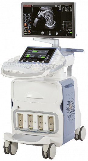
Voluson E10
Ultrasound scanner
Voluson E10 is an innovative ultrasound system from General Electric, which has collected all the advanced modern technologies. It helps obstetricians and gynecologists around the world to achieve the best image quality. The ultrasound scanner is fully digital and provides information to the specialist with a minimum of distortion.
The modern high-tech RSA (R-radiance S-System A-Architecture) platform was used as the fundamental basis on which the Voluson E10 is built. In addition, the system supports a new technology of matrix, volumetric electronic sensors. They allow you to get a high-definition image.

Volumetric visualization of blood flow and measurements in real time
Voluson E10 ultrasound system is a high-performance system focused on RealTime4D mode allows real-time reconstruction of volumetric images of anatomy and vessels in Doppler mode. The device allows using expert methods in the fields of:
- Reproductive medicine for visualization of follicles, fallopian tubes and embryo transfer;
- Oncology for examination of the endometrium and ovaries, microvascularization;
- Obstetrics for determining the tactics of childbirth;
- Fetal biometrics, diagnosis of heart defects, planning of invasive procedures;
- Gynecology for examination of uterine anomalies, pelvic floor muscles.

- Standard equipment of the device:
A 12-inch capacitive touch screen ensures sensitivity to touch and ease of control of the system; - A 22” OLED monitor with line-by-line scanning, high resolution;
- A control panel with the ability to change position;
- A 500 gigabyte hard drive;
- DVD+/-R(W) – a drive with the ability to record information on a DVD disc;
- 4 active ports;
- Intuitive user interface with the ability to ergonomically use the device menu using gestures;
- The device software is completely Russified.
Basic software modules:
- Presets for individual types of studies with the ability to reprogram, as well as a data storage/management system.
- HD-flow or so-called highly sensitive tissue Doppler.
- CRI is a complex scanning mode using several beams.
- XTD View is a panoramic image scanning.
- ATO is a system for automatic optimization of the image displayed on the display (works in any mode).
- SRI is a mode for suppressing various noises and artifacts (organ-specific).
- The standard HD-live program, which allows visualization of the fetus in volume, with an additional HDlive Silhouette application, including the Silhouette mode, with which you can highlight tissue boundaries and object contours. This package also includes the HDlive Flow mode, which is a light source that can be moved and is compatible with the blood flow visualization mode in volume.
- FFC (F-focus F-frequency C-composite) is the ability to multi-focus processing of the received signal.
- CE (C-coded E-excitation) – sending a pulse signal in coded form.
Inversion mode + 3D program (requires special sensors). - ScanAssistant – an application created for the convenience of conducting research; Special calculation program with the ability to create reporting documentation.
- Possibility of visualizing blood flow in B-Flow (non-Doppler mode).
SonoNT – a mode in which the calculation actions of the collar zone space are performed automatically; SonoIT – automatic calculation of the thickness of the 4th ventricle. - SonoBiometry – a mode that simplifies the measurement of such indicators (fetometric): BPD, OG, OB, DB, DP.
- 3D SonoRenderLive mode is an innovative system that allows you to determine the clear boundaries of the face and limbs of the fetus in volume, while getting rid of unnecessary artifacts, noise and interference.
- RePro is a function that enables the device to automatically connect the sensors required for the examination, as well as use all the necessary scanning parameters in order to reconstruct the conditions for the purpose of dynamic patient observation.
 392, N. Kapparov str., Almaty
 Mon-Fri 08:00-17:00 | Sat 08:00-13:00

 WhatsApp
 Instagram